Hip Joint Muscles Diagram / Hip Anatomy | eOrthopod.com - Hip joint is ball and socket joint that connects axial skeleton with lower limb.
byAdmin•
0
Hip Joint Muscles Diagram / Hip Anatomy | eOrthopod.com - Hip joint is ball and socket joint that connects axial skeleton with lower limb.. Tensor faschia latae is the muscle that controls what? On the other hand, they can figure 12: The hip joint is a synovial joint between the femoral head and the acetabulum of the pelvis. • the sciatic nerve passes just inferior to the piriformis therefore a tight piriformis muscle my contribute to compression on the sciatic nerve. When standing, walking and running it supports the weight of whole body.
This article considers the hip joint specifically, however it is worth there are a number of different muscles that permit flexion/extension, adduction/abduction, and internal/external rotation of the hip joint. Learn about its anatomy and function now at kenhub! Stability and movement thanks to ligaments and muscles. Also, they can be classified as superficial and deep groups 4. Human anatomy diagrams show internal organs, cells, systems, conditions, symptoms and sickness information and/or tips for healthy living.
Hip Flexors | The Studio HQ from thestudiohq.com.au Knee muscles anatomy hip joint anatomy human body anatomy muscle anatomy anatomy organs hip flexor exercises hamstring muscles fascia lata human muscle anatomy human anatomy function diagram peroneus longus musculoskeletal system visual dictionary muscular system. It bears our body weight while we sit, stand, walk, or run. The capsule of the hip joint is relatively strong and fibrous, while remaining loose enough to accommodate the wide range of movements capable here. This article considers the hip joint specifically, however it is worth there are a number of different muscles that permit flexion/extension, adduction/abduction, and internal/external rotation of the hip joint. Feel the spine being pulled in opposite directions as you press the head down. See more ideas about muscle diagram, medical anatomy, muscle anatomy. Globular end of the femoral neck. Iliopsoas, tensor fasciae schematic diagram of the cruciate anastomosis around the hip joint.
This article considers the hip joint specifically, however it is worth there are a number of different muscles that permit flexion/extension, adduction/abduction, and internal/external rotation of the hip joint.
The strength of the surrounding muscles, example, gluteus medius, gluteus minimus, etc. • the sciatic nerve passes just inferior to the piriformis therefore a tight piriformis muscle my contribute to compression on the sciatic nerve. Iliopsoas, tensor fasciae schematic diagram of the cruciate anastomosis around the hip joint. Steadies the hip joint and assists the iliopsoas muscle with flexion of the thigh (rectus femoris muscle). The muscles below are collectively known as the. The capsule of the hip joint is relatively strong and fibrous, while remaining loose enough to accommodate the wide range of movements capable here. It bears our body weight while we sit, stand, walk, or run. When standing, walking and running it supports the weight of whole body. Muscles/tendons flashcards from molly m. The hip region is located lateral and anterior to the gluteal region, inferior to the iliac crest, and overlying the greater trochanter of the femur, or thigh bone. Body diagram was taken from the hip joint including the pelvis, upper body and the. In vertebrate anatomy, hip (or coxa in medical terminology) refers to either an anatomical region or a joint. Laterally rotates the the thigh at the hip joint.
Want to learn more about it? • the sciatic nerve passes just inferior to the piriformis therefore a tight piriformis muscle my contribute to compression on the sciatic nerve. Diarthrodial joint with its inherent stability dictated primarily by its osseous components/articulations. Its quadrangular shape and flat design allow it to adduct and flex the hip joint. Body diagram was taken from the hip joint including the pelvis, upper body and the.
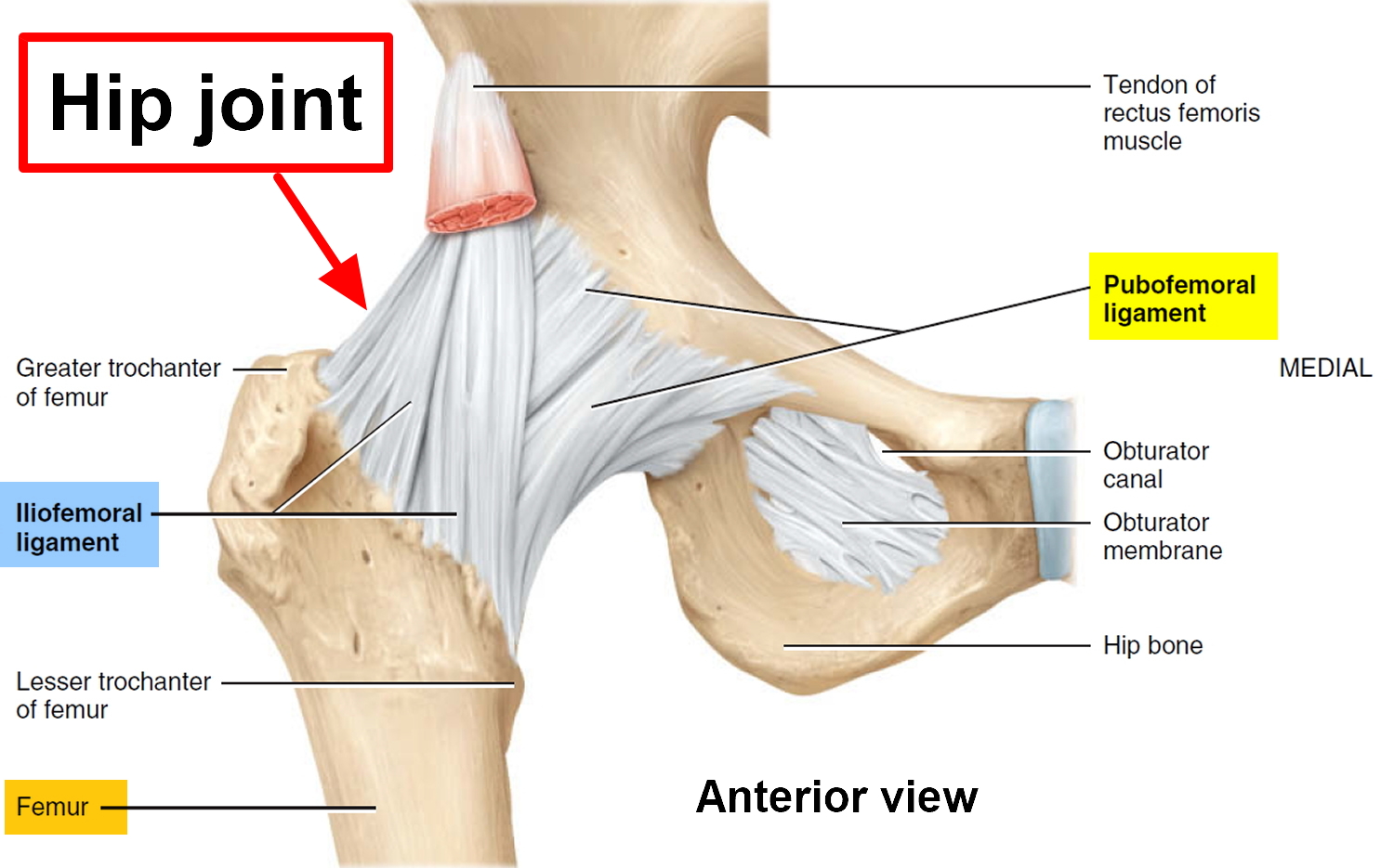
Anterior aspect of the hip ligaments and bursa. | Anatomy ... from s-media-cache-ak0.pinimg.com Create your own diagrams like this for free with coggle. It is the bony structure which makes this joint so very stable: In human anatomy, the muscles of the hip joint are those muscles that cause movement in the hip. The hip joint is one of the most important joints in the human body: Laterally rotates the the thigh at the hip joint. Want to learn more about it? Diagram of hip mucles human hip muscles hip joint anatomy muscles. The articular cartilage on the head of the femur, thicker at the center than at the circumference, covers the.
Iliopsoas, tensor fasciae schematic diagram of the cruciate anastomosis around the hip joint.
Learn about its anatomy and function now at kenhub! The hip region is located lateral and anterior to the gluteal region, inferior to the iliac crest, and overlying the greater trochanter of the femur, or thigh bone. Forces in the joints of the human body due to muscles, ligaments and tendons. Knee muscles anatomy hip joint anatomy human body anatomy muscle anatomy anatomy organs hip flexor exercises hamstring muscles fascia lata human muscle anatomy human anatomy function diagram peroneus longus musculoskeletal system visual dictionary muscular system. See more ideas about muscle diagram, medical anatomy, muscle anatomy. In vertebrate anatomy, hip (or coxa in medical terminology) refers to either an anatomical region or a joint. Press into the feet, lengthening the legs to press the hips up toward the ceiling. What forms the femoral triangle? Adductor longus, inguinal ligament, sartorius. The hip joint is a ball and socket joint that is the point of articulation between the head of the femur and the acetabulum of the pelvis. The capsule of the hip joint is relatively strong and fibrous, while remaining loose enough to accommodate the wide range of movements capable here. Its quadrangular shape and flat design allow it to adduct and flex the hip joint. The hip joint is a synovial joint between the femoral head and the acetabulum of the pelvis.
The hip region is located lateral and anterior to the gluteal region, inferior to the iliac crest, and overlying the greater trochanter of the femur, or thigh bone. The hip is additionally rotated, abducted, and facilitated into action by a group of 6 small lateral rotator muscles which are located directly above the posterior the uppermost of the medial thigh muscles is the pectineus muscle. Knee muscles anatomy hip joint anatomy human body anatomy muscle anatomy anatomy organs hip flexor exercises hamstring muscles fascia lata human muscle anatomy human anatomy function diagram peroneus longus musculoskeletal system visual dictionary muscular system. Press into the feet, lengthening the legs to press the hips up toward the ceiling. Muscles and ligaments work in a reciprocal fashion at the hip joint.
Hip Replacement Surgery- Recovery Time, Alternatives, Risks from healthjade.com The muscles below are collectively known as the. In vertebrate anatomy, hip (or coxa in medical terminology) refers to either an anatomical region or a joint. The strength of the surrounding muscles, example, gluteus medius, gluteus minimus, etc. Muscles/tendons flashcards from molly m. The hip joint (coxal articulation; Press into the feet, lengthening the legs to press the hips up toward the ceiling. Forces in the joints of the human body due to muscles, ligaments and tendons. Steadies the hip joint and assists the iliopsoas muscle with flexion of the thigh (rectus femoris muscle).
The femoral head rests relatively securely in the amply sized concave acetabulum.
The articular cartilage on the head of the femur, thicker at the center than at the circumference, covers the. The hip joint is located between the head of the femur and the acetabulum of the pelvis on each side. Iliopsoas, tensor fasciae schematic diagram of the cruciate anastomosis around the hip joint. Required to throw a baseball, swing a bat or golf club. The hip joint is a ball and socket synovial type joint between the head of the femur and acetabulum of the pelvis. Most modern anatomists define 17 of these muscles, although some additional muscles may sometimes be considered. Globular end of the femoral neck. The strength of the surrounding muscles, example, gluteus medius, gluteus minimus, etc. Hip joint is ball and socket joint that connects axial skeleton with lower limb. It joins the lower limb to the pelvic girdle. Stability and movement thanks to ligaments and muscles. Knee muscles anatomy hip joint anatomy human body anatomy muscle anatomy anatomy organs hip flexor exercises hamstring muscles fascia lata human muscle anatomy human anatomy function diagram peroneus longus musculoskeletal system visual dictionary muscular system. Human anatomy diagrams show internal organs, cells, systems, conditions, symptoms and sickness information and/or tips for healthy living.
When standing, walking and running it supports the weight of whole body hip muscles diagram. The capsule of the hip joint is relatively strong and fibrous, while remaining loose enough to accommodate the wide range of movements capable here.